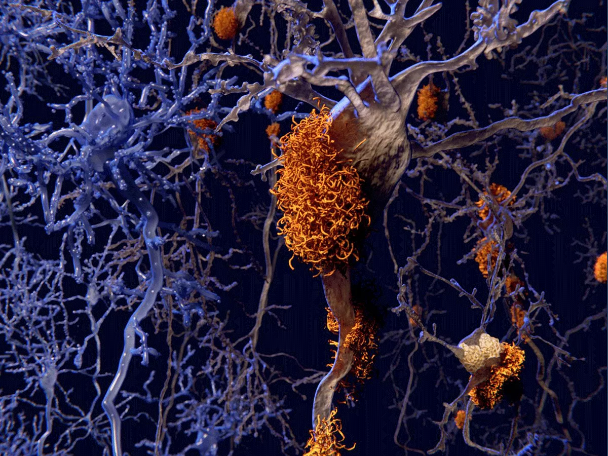
Tau protein aggregates are present in a group of disorders, collectively termed the tauopathies. Alzheimer's disease (AD) is the most common of those disorders, while frontotemporal dementia (FTD) is most strongly linked to tau.
Now, a map of the proteins that interact with tau and how those interactions differ between normal and disease-associated tau protein could give new clues on how the protein causes damage in neurodegenerative disorders, and on how to treat or prevent that damage.
Investigators published their work in the January 20, 2022, issue of Cell.
Unlike amyloid-beta, which is an extracellular protein, tau is found inside cells. It plays important roles in stabilizing microtubules, which are important for protein transport -- its full name is microtubule-associated protein tau (MAPT).
Neurons are particularly dependent on a functioning transport infrastructure. Neuronal connections can bridge long distances, and in the most extreme cases, proteins can travel several feet from their production site in the nucleus to the synapses.
But tau is also unusual because it spreads from neuron to neuron as tauopathies progress, and "there is the possibility that interfering with spreading could slow progression," Tara Tracy told BioWorld Science.
Tracy is an assistant professor at the Buck Institute for Research on Aging, and the paper's corresponding author.
In their work, Tracy and her colleagues used iPSCs to generate neurons and determine tau's interaction partners under several different conditions.
Previous work had shown that tau spread when neurons were active, and so the researchers looked at the binding of wild-type tau both when neurons were resting, and when they were firing.
They also compared the binding behavior of normal tau to two versions of the protein with FTD-associated mutations, TauP301L and TauV337M.
They showed that in active neurons, tau associated with a protein called synaptotagmin, which is part of a protein complex called SNARE that orchestrates the release of neurotransmitter packets.
"That suggests that tau could get secreted from neurons through the mechanism that allows vesicles to be released from neurons," Tracy said.
The team also showed that "tau associated with multiple mitochondrial proteins, which is very interesting," she said. "Which ones would be important to target is something we don't know yet, but which could be really interesting to pursue" from a translational standpoint.
The team also looked at the expression of those mitochondrial proteins in postmortem tissue of patients with different tauopathies, including AD, mild cognitive impairment, frontotemporal dementia and progressive supranuclear palsy. They found that expression of tau-interacting mitochondrial proteins was reduced in those tissues, and that lower expression levels were associated with more severe disease in several conditions.
The authors wrote in their paper that "these data suggest that the [tau]-mitochondrial interactors identified in vitro using human neurons are downregulated in both primary and secondary tauopathies and are associated with both cognitive decline and the accumulation of pathological tau in the brain."
"Translationally," Tracy summarized, "you would want tau to stay bound."
Which tau, exactly?
The final protein aggregates of tau that are the most easily visible sign of tauopathy may not actually be the most problematic ones. Aggregates form when peptides assemble into protofilaments, which assemble into filaments, which assemble into fibrils, which aggregate.
The cause of pathology is "not a clear picture, because there are a lot of forms that could play a role," Tracy acknowledged. And for tau, the picture is even more complicated because it is heavily phosphorylated. "That adds complexity -- how do all these modifications cause problems."
At this point, she said, there is "general consensus that the big aggregates are actually not what's causing toxicity in the brain."
Instead, the problem seems to be fibrils and small oligomers, and perhaps non-aggregated monomers.
"We think that our study primarily involves either monomers, or maybe small aggregates -- we actually don't know," she said. "I can't say whether they are fibrils in our system or not."
For this reason, the team may not be identifying some additional proteins that are interacting with fibril rather than monomer tau.
However, fibrillar tau is unlikely to be a major binding partner for other proteins.
"I'm just speculating here... we don't have evidence for that, because we didn't design the study that way," Tracy stressed. But fibrillar tau could be "less likely to interact with other proteins, [because] fibrillar tau is a whole lot of tau proteins interacting with each other."
