MUNICH, Germany – The 2008 congress of the European Society of Cardiology (ESC; Sophia Antipolis, France), which ended here in early September with record attendance in excess of 30,000, is the leading venue for introduction of new cardiovascular device technologies in Europe, and for presentation of clinical findings in European cardiology.
The focus of this year's congress was on cardiovascular imaging, which covers a wide range of non-invasive imaging modalities including CT, MRI, echocardiography, SPECT and PET imaging. Invasive imaging technologies such as intravascular ultrasound (IVUS) and, on an experimental basis, optical coherence tomography (OCT), also are being used increasingly in the diagnosis and management of cardiovascular disease in Europe, and advances are occurring in invasive angiography as well.
Imaging products represented a $30 billion worldwide market in 2007, as shown in Table 1, growing at almost 7% per year. Cardiovascular applications account for a significant proportion of imaging procedures, particularly in the ultrasound and nuclear imaging segments, and comprise a growing proportion of procedures in CT and MR. Hybrid imaging, combining technologies such as CT and SPECT or CT and PET, is also becoming increasingly important in cardiology, enabling more rapid and comprehensive assessment of cardiovascular disease to be performed with greater ease, improving the efficiency of patient management. Integration of imaging technologies is expanding, based on new developments reported at the ESC congress, to include CT/ultrasound, CT/MR, MR/PET and angiography/IVUS hybrid imaging.
 |
An important new theme emerging at the ESC congress is imaging of ischemia, as opposed to imaging of anatomy, the latter being the role of conventional coronary angiography, the current gold standard for diagnosis of coronary artery disease and guidance of interventional therapy. Ischemia is assessed via nuclear imaging (SPECT) in most patients now, with other techniques such as ECG and invasive fractional flow reserve (FFR), a parameter measured using the PressureWire from Radi Medical Systems (Uppsala, Sweden), also playing a role. Cardiologists are seeking improved methods for disease assessment since anatomy does not always indicate the presence of ischemic tissue, while perfusion measurements such as SPECT imaging can miss some patients who have significant coronary artery disease. Other areas highlighted at the ESC congress included the latest developments in the treatment of heart failure, advances in prevention of cardiovascular disease, the status of drug-eluting stents, and recent developments in cardiac rhythm therapy.
Advances on multiple fronts
CT is becoming an increasingly important modality for cardiovascular imaging as a result of technological advances that have led to higher resolution, more rapid scan times, and more powerful image processing capabilities. The most recent advances have enabled radiation dose to be reduced considerably, addressing one of the primary drawbacks of coronary CT imaging.
Toshiba Medical Systems (Tokyo) is a leader in the segment with its Aquilion ONE system, introduced in November 2007. The Aquilion ONE is the first 320-slice CT scanner, and provides a significant increase in scan density compared to the latest-generation 64-slice scanners.
Toshiba has recently added the capability to perform alternating scans at a 0.25 mm offset, providing an effective 640-slice scan via image reconstruction. The Aquilion ONE also uses a 16 cm wide area detector, providing a resolution of 0.5 mm, with an image acquisition time or temporal resolution of 350 ms. Those features enable users to image the coronary arteries non-invasively to detect stenoses with sensitivity approaching that of invasive angiography.
Current-generation 64-slice scanners usually over-estimate the degree of stenosis, resulting in a high false positive rate when performing population screening via CT. The company has also developed the ConXact image reconstruction software which reduces blooming of the image in the presence of calcium deposits, enabling more accurate detection of stenosis.
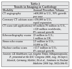
The use of CT to screen individuals who have no symptoms of heart disease has expanded rapidly. As shown in Table 2, more than 150,000 coronary CT scans are performed annually in the U.S. to obtain a coronary calcium score, which quantitates the degree of calcification or hardening of the arteries without the use of a contrast agent. Many patients in hospitals in Europe request such exams, according to experts presenting at the congress.
 |
Coronary CT can be an effective test in populations with a low to moderate risk of coronary artery disease, as discussed by Fabio Esteres, MD, of Emory University (Atlanta), at the ESC congress. The exam costs about $200, and can be a cost-effective means to select patients for additional, more expensive diagnostic tests. In patients without chest pain who have a negative coronary calcium score, only one in 200 will die within 10 years of heart disease, while in patients with chest pain a negative score predicts absence of coronary artery disease in 99 out of 100 patients.
Patients with a positive score can have a SPECT or PET test to further stratify their disease risk and determine the need for treatment. Since a PET exam costs $1,700, the strategy can reduce overall cost by almost one-third compared to using PET alone. In a study performed at Emory by Esteves, the savings amounted to $617 per patient.
CT angiography (CTA), which adds the use of contrast to the CT imaging procedure, is also being used in a growing number of patients worldwide as a non-invasive approach to risk stratification. As shown in Table 2, the procedure is performed in over 400,000 patients in the U.S., and based on the ratio of total CT procedures in the U.S. to worldwide procedures, at least twice that number are performed worldwide.
The primary role of CT angiography in cardiology at present, as discussed by Joseph Schuijf, MD, of Leiden University Medical Center in the Netherlands, is in ruling out coronary artery disease in patients with chest pain. CT angiography has a very high (more than 90%) negative predictive value for coronary artery disease detection. Because a CT angiogram can be performed rapidly and at a relatively low cost, it can be used in patients without conclusive signs of a heart attack.
In the Core 64 sub-study, presented at the ESC congress by Carlos Rochitte, MD, of the University of Sao Paulo Medical School in Brazil, multi-detector CT was shown to have equivalent high sensitivity for detection of coronary stenosis in both ACS and non-ACS patients. With the latest-generation CT systems, spatial resolution is approaching that of invasive angiography. For example, the newest unit from GE Healthcare (Chalfont St. Giles, UK), the CT750 HD, has a resolution of 0.23 mm and acquires 7,000 image slices during each rotation.
At that level of resolution, the system is capable of imaging stents, localized plaque characteristics, and degree of stenosis with a precision approaching that of conventional angiography, which has a spatial resolution of 0.2 mm. Image acquisition time/temporal resolution for the CT750HD, which can be as short as 65 ms using the SnapShot Burst Plus feature, still lags that for conventional angiography of about 8 ms, however, so image artifacts related to vessel motion may still limit the accuracy of CTA relative to angiography.
A major issue for CT, however, is radiation exposure. As the use of CT scans has expanded, including scans performed in individuals without symptoms of disease, concerns have arisen about the risk of inducing cancer in individuals exposed to increasingly large cumulative radiation doses. The radiation dose for a single CT exam using scanners available prior to the latest-generation machines was typically about 15 millisieverts (mSv), a level that is about 100 times the dose received in a standard chest X-ray.
As discussed by Francesco Faletra of Fondazione Cardiocentro Ticino (Lugano, Switzerland), a 10 mSv CT study is estimated to be associated with an increase in risk of fatal cancer of one in 2000. Since many patients may undergo multiple scans, it has not been uncommon for a patient to receive 50 to 100 mSv in a relatively short period of time, according to presenters at the ESC congress.
However, manufacturers of CT systems have responded by introducing a new generation of scanners with significantly lower radiation dose characteristics. The new Aquilion One scanner from Toshiba, for example, can scan the entire heart in one beat, versus multiple (five or more) beats in prior generation scanners, resulting in radiation doses as low as 2 mSv per scan.
Furthermore, new image acquisition techniques such as ECG-gated imaging (Step-and-Shoot) enable additional reduction in dose, since the radiation source is only on for a small portion of the cardiac cycle. GE's new CT750HD scanner uses a prospective gating technique called SnapShot that results in an 83% reduction in dose, dropping exposure to a level of 3 mSv, and with additional planned refinements it may be possible to reduce the dose to 0.6 mSv per scan, less than the allowable annual dose for the general population of 1 mSv.
Siemens Medical (Munich, Germany), which has the largest installed base of 64-slice CT scanners worldwide with its Somatom Sensation, also is developing methods for dose reduction using ECG-gating technology. Radiation dose reduction is expected to be an important competitive focus for manufacturers of CT scanners going forward in order to reduce safety concerns as a barrier to more widespread use of the technology.
Molecular imaging using nuclear imaging technology is expected to become increasingly important in cardiology, according to a number of presenters at the ESC congress. At present, the number of PET and SPECT imaging procedures performed is the smallest of any of the major imaging modalities in cardiology, but total nuclear imaging procedures (PET and SPECT) are growing at 12% per year worldwide, according to suppliers.
The long-term goal, as discussed by Franck Bengel, MD, of Johns Hopkins Medical Institutions (Baltimore), is to use molecular imaging to detect in vivo biomarkers of cardiovascular disease, enabling earlier detection, personalized therapy, and more effective monitoring. Imaging methods such as CT and ultrasound mainly analyze morphology and physiology, whereas molecular imaging is able to characterize tissue at the subcellular level.
Combining morphological imaging methods such as CT with molecular imaging enables characterization of the complete disease process, according to Bengel. As an example, Bengel cited a recent animal study performed by his group in which PET imaging of tissue perfusion and catecholamine uptake were used to identify tissue regions that might be at risk for ventricular arrhythmia following an experimentally induced myocardial infarction.
The areas identified as having elevated risk by molecular imaging correlated with those showing an abnormality via subsequent invasive voltage mapping. The results indicate that molecular imaging could provide a non-invasive method to evaluate post-myocardial infarction patients to predict the risk of ventricular arrhthymia.
Another new technique for molecular imaging is under development by Siemens Medical that employs ECG-gated SPECT imaging to improve the ability to assess perfusion in segments of the coronary arteries.
Progress in MR imaging in cardiology was another focus at the ESC congress. The spatial resolution of MR is continuing to improve, although not yet to a level that allows it to compete in that respect with CT scans, but MR also has unique capabilities for tissue characterization including molecular imaging that may make it a more powerful modality in the future.
MR is proving particularly useful in detection of myocardial scars that result from an ischemic event, and which compromise the ability of the heart to function normally and to recover from a heart attack.
In some cases, as discussed by Peter Buser, MD, of University Hospital (Basel, Switzerland), cardiac MR can provide information that is equivalent to SPECT. However, its ability to characterize tissue such as scar and plaque at a high level of resolution, 40-fold better than SPECT, provides a potential advantage over nuclear imaging techniques. Myocardial tissue viability also can be assessed with cardiac MR, as can microvascular obstruction, which can be used to assess the ability to recover from a myocardial infarction.
A key advantage of MR vs. CT, conventional angiography, or nuclear imaging is its lack of radiation exposure for both the patient and the physician. At least one presenter at the ESC congress, Heiko Mahrholdt, MD, of Robert Bosch Medical Center (Stuttgart, Germany), believes cardiac MR will replace SPECT within the next decade as the preferred modality for evaluation of myocardial viability. Accuracy is equivalent to SPECT or echocardiography, and MR can be safely performed in patients with acute coronary syndromes, giving it a broader spectrum of application than nuclear techniques.
MR also is a powerful modality for detection of reperfusion following treatment of a coronary occlusion, and with the use of molecular MR imaging agents the technique is showing promise for ischemic memory imaging, which could prove useful in assessing the extent of myocardial infarction.
Leading suppliers of MR scanners include Siemens Medical, the market share leader in the segment; GE Healthcare; Philips Medical (Best, the Netherlands); and Toshiba Medical.
Advances in cardiac ultrasound
Echocardiography, or cardiac ultrasound, is the most widely used imaging modality in cardiology, as indicated by the data in Table 2. A revolutionary development in cardiac ultrasound was unveiled at the ESC congress by Siemens Medical, the new SC2000 ultrasound system, which has the capability to acquire a complete 3-D volumetric image of the heart in a single heartbeat. Previous-generation ultrasound systems require images from four to seven beats to create a single composite 3-D image. The SC2000 enables instantaneous imaging of cardiac volume, and allows cardiac ejection fraction to be measured in a single heartbeat.
The new technology should improve the ability to perform non-invasive measurements of cardiac parameters, since artifacts due to unstable heart rate are avoided. In addition, it enables more rapid measurements to be performed and is easier to use than prior-generation ultrasound imaging systems, eliminating the need for breath-hold or ECG gating. More accurate measurement of parameters such as the left ventricular function index and left ventricular injection fraction is needed in echocardiography, since changes in those parameters in disease states are typically only 3-4%, while the variability in measurement with prior-generation ultrasound systems is 7% to 8%.
GE Healthcare also introduced a single-heartbeat 4-D ultrasound system, the Vivid E9, at the ESC congress. Another advance is improvements in transthoracic echo, which in combination with CT scanning allows a high percentage (98%) of coronary arteries to be imaged non-invasively, i.e., without resorting to invasive angiography, enabling both vessel anatomy and blood flow to be analyzed in a single fused image.
Real-time 3-D transesophageal echo, introduced in 2007 by Philips Medical as the iE33 ultrasound system, is generating strong interest among cardiologists for applications in mitral valve surgery guidance and to guide various heart defect repair procedures such as closure of PFOs and atrial septal defects.
Numerous presentations at the ESC congress focused on the use of cardiac ultrasound to evaluate patients being considered for treatment with implantable cardiac rhythm therapy (CRT) devices. At present, diagnostic techniques used to select patients for CRT implants are far from perfect: 30% of all patients receiving implants do not show a clinical response to therapy, 40% fail to show improved left ventricular function, and for a number of patients receiving ICDs the implant proves unnecessary since it never fires.
A wide variety of new techniques have been evaluated to improve patient selection, including MR imaging to assess myocardial scar, but so far none have improved patient outcome and selection. At ESC, George Sutherland, MD, of St. George's Hospital (London), discussed the use of cardiac ultrasound for patient selection in CRT. Sutherland has found that presence of an ultrasound image feature called septal flash, which can easily be discerned by visual inspection of an image, is highly predictive of response to CRT.
Septal flash also can be measured via tissue Doppler, 2-D strain imaging and M-mode ultrasound. If CRT is implemented in a patient exhibiting septal flash, the image feature disappears, and the patient responds both with an improvement in cardiac volume as well as in clinical factors.
Sutherland has found that five groups of patients exist, four of which exhibit certain features on cardiac ultrasound and respond to CRT and one group which does not exhibit such features and does not respond. In addition to septal flash, responder groups are identified by the presence of long atrio-ventricular delay, short atrio-ventricular delay, and right ventricle-left ventricle delay. The measurements are performed using stress echocardiography with a dobutamine challenge.
Sutherland's group now tests all patients scheduled for CRT with dobutamine stress echo for presence of septal flash and other parameters predictive of response.
Diastolic dyssynchrony, another parameter that can be measured via cardiac ultrasound, also was discussed at the ESC meeting as a potential metric for prediction of CRT response. Anthony DeMaria, MD, of San Diego, editor of the Journal of the American College of Cardiology, in a presentation on recent developments in cardiac ultrasound, stated that diastolic dyssynchrony has become more important for management of heart failure patients in the past year.
The PROSPECT (Predictors of Response to CRT) trial assessed the use of diastolic dyssynchrony to predict the benefit of CRT. However, the results of PROSPECT showed that only two-thirds of patients selected by diastolic dyssynchrony benefited, and Receiver Operating Characteristic (ROC) curve analysis showed the parameter was not significantly better than random chance in guiding patient selection.
While improvement in the reproducibility and accuracy of echocardiography measurements, as well as reduction in operator dependence, could potentially product better results, experts presenting at ESC said that no ultrasound measurement currently has a role in CRT therapy selection. That conclusion, based primarily on the results of PROSPECT with diastolic dyssynchrony, makes the data from Sutherland's group even more interesting as a potentially more powerful method for CRT selection.
A number of companies introduced new products for cardiac ultrasound at the meeting. Philips Medical launched the CX50 CompactXtreme hand-carried ultrasound system, which has similar performance to the iE33 system from Philips but in a portable configuration. The CX50 is priced similar to the Vivid i from GE Healthcare. The system does not yet incorporate the real-time 3-D capabilities of the iE33, but plans call for those features to be added.
Esaote (Florence, Italy) showed the new MyLab 30 Gold, an upgrade of the MyLab 30 CV portable ultrasound system that provides 100 minutes of battery-driven operation, and Kontron Medical (Plaisir, France) introduced the ImagicAgile portable ultrasound system.
GE Healthcare launched the Vivid q, a new portable ultrasound system employing active matrix array technology which can perform automated measurement of ejection fraction and intra-cardiac echo.
Portable ultrasound systems are now a major category in the ultrasound market in Europe, along with mid-level and high-end fixed-base systems. Some suppliers expect the market to consolidate into only two segments, portables (including handheld and hand-carried systems) and high-end fixed-base systems. Portables are in demand among private cardiologists in office-based practices in Europe, and also are preferred by many users in clinics and hospitals because they occupy less space, emit less noise, and provide flexibility.
Ultrasound may eventually become an even more ubiquitous modality, based on emerging approaches described at the ESC gathering. For example, a development-stage device for bedside ultrasound monitoring of pressure displacement loop as an indicator of post-procedural myocardial ischemia was described by Thor Edvardsen and others from (Rikshospitalet University Hospital (Oslo, Norway). The device is being developed in collaboration with Imasonic (Besancon, France), and consists of a miniaturized ultrasound transducer that is implanted in an epicardial position, similar to a temporary pacemaker lead. It allows analysis of M-mode and flow velocities to estimate vessel wall characteristics correlated with ischemia.
A separate arterial blood pressure measurement is required which in the clinical situation would simply be provided by the pressure transducer normally attached to the patient's arterial catheter. The device is being evaluated in animal studies. The primary application envisioned is for continuous monitoring for ischemia in patients following a CABG procedure, since ECG is not sufficiently sensitive and specific to reliably detect such events.
Growing use of remote monitoring
Another area highlighted at the ESC congress was remote patient monitoring, a modality that is being used increasingly for disease management in Europe, particularly for patients with heart failure. A number of technologies are being employed. The most widely used monitoring technology is implantable monitors, most of which are incorporated into cardiac rhythm control devices such as pacemakers and ICDs.
As shown in Table 3, leaders in the worldwide implantable device monitoring segment include Medtronic (Minneapolis), Biotronik (Berlin, Germany), Boston Scientific (Natick, Massachusetts) and St. Jude Medical (St. Paul, Minnesota).
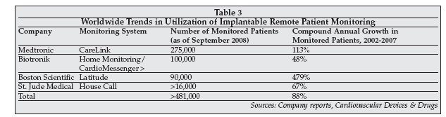 |
The number of patients being monitored with implantable cardiac rhythm device monitoring systems is projected at in excess of 500,000 at year-end 2008. Growth in the number of monitored patients is estimated at 88% per year over the 2002-2007 interval.
At present, the Medtronic CareLink network monitors patients mainly in the U.S. and Europe, while about 40% of the patients monitored by the Biotronik Home Monitoring system are in the U.S. and 50% are in Europe. Both the Guidant Latitude and the St. Jude HouseCall systems are deployed primarily in the U.S. Introduction of the Latitude system in Europe is planned for 1Q or 2Q09.
The systems are designed to track device performance, such as the number of shocks delivered by an ICD, as well as device failures such as battery depletion or lead failure. They also can track the patient's ECG and heart rate, and with additional external sensors parameters such as body weight, temperature, and blood pressure can be remotely monitored.
Data from the implanted device is transmitted wirelessly to a remote monitor in the patient's home, either automatically or via a sequence initiated by the patient. Once downloaded, the data can be sent to a central server via telephone modem, where it is analyzed by the host computer.
Alerts are generated if a device fault is detected or if patient parameters deviate from prescribed norms, and a clinician, typically an EP nurse, takes appropriate action. Cardiologists also can access the data on each patient via the Internet.
At the ESC congress, Biotronik previewed its next-generation Home Monitoring system, which will be launched at the end of March 2009. The new system addresses a number of issues discussed by cardiologists at the congress that have arisen with current-generation systems. For example, many hospitals are now overloaded with data from implantable monitors. Data is typically transmitted daily for each monitored patient, or more often if an alert situation occurs.
As the number of monitored patients has expanded, the volume of data has grown to a level that makes it impossible for a nurse to review all records on a daily basis. Instead, the central server software automatically analyzes the data and generates a simplified summary along with alerts.
In the case of the new Biotronik monitoring system, the user interface will be further simplified, adding color-coding of events (yellow for important events, red for serious events).
In Europe, remote monitoring is now considered equivalent to in-person follow-up once the patient enters the stability period (two months post-implant), and the Heart Rhythm Society (Washington) now recommends the use of remote monitoring in all ICD patients.
The Biotronik monitoring system operates worldwide, using the cellular telephone network to transmit data to the monitoring center in Germany. Cost of monitoring is included in the price of the implantable device in the case of the Biotronik system, while for other systems there may be an additional fee for the service.
A number of other remote monitoring technologies were described at the congress which can be used on patients who do not have implanted devices. Josiane Janssen-Boyne, a nurse-practitioner in a group at Maastricht University Hospital (Maastricht, the Netherlands) described a trial of the Health Buddy home monitoring system from Health Hero Network (Palo Alto, California) being conducted at three Dutch centers.
The Health Buddy is a patient interface device that can acquire blood pressure, spirometry, weight and blood glucose data from external devices and also allows information to be input via pushbutton responses to a series of scripted questions. The data is transmitted via telephone modem to a monitoring center.
In the Netherlands trial, patients were also contacted by telephone to discuss their health status and any recommended changes in treatment. Initial results from the trial indicate a significant improvement in quality of life and therapy compliance, as well as an improved level of understanding by the patients of their medical condition. The study also found that monitored patients had a high incidence of clinical depression, but Janssen-Boyne said that finding may be related to the time period over which data from the study is currently available, which includes the winter months.
Health Hero has achieved significant penetration in the home telehealth market, with more than 20,000 individuals with chronic health conditions currently being monitored worldwide. The company is seeking to expand its penetration in regions outside the U.S., including Europe.
Results of a study of home telemonitoring for management of heart failure using another leading telemonitoring system, the Genesis monitor from Honeywell HomMed (Brookfield, Wisconsin), were presented at ESC by Jillian Riley of Imperial College (London). At 120-day follow-up in the HOME-HF study, which was conducted in a group of 182 patients discharged from three London hospitals, there was no difference in repeat hospitalization between the monitored and the usual care groups. However, the telemonitored patients had fewer unplanned hospitalizations and few outpatient visits.
In discussing the results in the highlight session for the ESC congress, Helmut Drexler, MD, of Medizinische Hochschule Hannover in Germany, speculated that the lack of reduction in hospitalizations could be due to more intensive care in the telemonitored group during the initial period following discharge.
Aipermon (Munich, Germany) exhibited its remote monitoring system, which can employ either of two monitoring devices, the AiperCoach Mobile Medical Assistant (MMA) or AiperCoach Home (Homebox). The MMA is a PDA device that acquires physiological data wirelessly from various sensors (e.g., a wrist blood pressure meter) and also provides two-way communication between the patient and a monitoring nurse or physician to check medication compliance and clinical status. Monitored parameters include ECG, physical activity, temperature, blood pressure, body weight, and spirometry.
The Homebox is a fixed-base station that is located in the patient's home to acquire monitoring data and transmit it to the Aipermon Data Service Center. The company has a worldwide contract with Verizon Communications (New York) for data transmission, and either rents or sells its system to hospitals and other users such as companies which perform clinical trials. The MMA or the Homebox are purchased at a cost of 300, or rented for a rate of between 8 and 25 depending on the number and type of measurement devices used.
At present, the Aipermon system is deployed only in Europe, with 10 to 15 hospitals and other facilities now using the product. Major applications for the Aipermon telemonitoring system include management of congestive heart failure and weight loss programs (via activity monitoring). A recent study found that telemonitoring with the Aipermon system resulted in a tripling of weight reduction in monitored patients compared to a control group.
Apoplex Medical Technologies (Pirmasens, Germany) exhibited the SRAclinic, a stroke risk analysis system, at the congress. SRAclinic analyzes 24-hour ECG data using proprietary mathematical methods based on assessment of R-dynamics to detect paroxysmal atrial fibrillation, even if the AF event occurred prior to the test.
SRAclinic is used primarily in hospital stroke units for secondary prevention, and can be used on an on-going basis to monitor patients following a stroke to assess risk for recurrence. The system requires an Internet connection, since the ECG data is transmitted to Apoplex for analysis, with the results returned to the hospital within a few minutes. Between 200 and 250 general practitioners as well as 10 stroke clinics in Germany are using the Apoplex SRAclinic.
HealthSTATS International (Singapore) exhibited the BPro Radial Pulse Wave Acquisition Device. BPro uses a proprietary technology, EVBP, to measure arterial blood pressure waveforms using an external radial artery pressure transducer. In conjunction with the company's A-Pulse CASP pulse wave analysis software, the BPro can capture and display the radial artery pulse wave, and can derive a number of parameters such as central aortic systolic pressure, radial augmentation index, and a range of other parameters that can be used to assess arterial stiffness and cardiovascular health.
With the BProSOFT software, 24-hour ambulatory central blood pressure monitoring can be performed. Studies have shown a correlation of greater than 0.99 between central blood pressure derived from the BPro and directly measured central pressure.
HealthSTATS also is developing a new version of the BPro that will have applications in assessment of stroke risk based on non-invasive measurement of blood pressure in the superficial temporal arteries