By MICHAEL SIMONSEN
Cardiovascular Device Update Contributing Editor
and DON LONG
Cardiovascular Device Update Executive Editor
ORLANDO, Florida — Let's start with an obvious fact.
Over the past five years significant advances have marched out of the laboratories and clinical trial factories of the cardiovascular disease sector.
These advances have consisted of three major developments: new drugs, new devices and new diagnostics. And of these three, devices have led the way in innovation, in the ability to add to years of quality life, in major technology breakthroughs, in some of the largest headlines — good and bad — and in offering the most controversy.
Some might argue that drugs have led the way, but you certainly didn't see this very well supported at this year's scientific sessions of the American Heart Association (Dallas) in early November. The sessions dealing with drugs often seemed like beating the proverbial "dead horse," since dealing with the continuing concerns of drugs to lower cholesterol, and more often, ongoing debates about the appropriate uses of blood thinners and anti-platelet therapies — this latter largely device-driven since putting a large and bright spotlight on stent implantation and how to avoid thrombosis and other adverse events.
Diagnostics in the sector is serving to better analyze the heart disease you are experiencing now, but it still hasn't come close to solving the most important "gold standard" questions: the ability to predict serious oncoming cardiac events — such as stroke and aneurysms — among asymptomatic patients; or the ability to improve the dismal predictive quality of tests that attempt to determine who will actually benefit from an implantable cardioverter defibrillator (ICD) — currently at less than 10%.
Devices recently have clearly outperformed drugs and diagnostics in terms of products of really innovative and market-changing dynamics (DES and peripheral stenting) as well as clinical benefit (artificial hearts and ventricular pump devices that are increasingly be shown as able to "remodel" the heart, and on the horizon new implantable systems for stroke therapy).
And devices have created major headlines with recalls of defective pacing systems (and most recently the leads for those systems). And the device world has created the story with the most legs: the debates concerning stent use — vs. medical therapy, vs. coronary bypass procedures and the actual numbers of post-implant risks for DES. These debates are not likely to end soon.
Theme: technology …
and more technology
Thus, the AHA sessions were the perfect venue oforthe array of new developments in therapeutic devices — as well as drugs and diagnostics. And its over-arching theme was the rapidly increasing role in the use of technology for management of the disease, and systems for aiding patients in disease management.
New evidence on the safety of the current generation of drug-eluting stents was presented, and also on next-generation stents, coupled with advances in anti-thrombotic drug therapy, which promise to substantially reduce, if not eliminate, the drawbacks of present-generation devices.
Advances in percutaneous repair of heart valves were also described, as well as emerging technologies for treatment of cardiac arrhythmias that show promise for improving the success rate of cardiac ablation procedures and, potentially, enabling non-invasive treatment which could allow a broader range of patients to be addressed.
In the diagnostics arena, there is a growing trend toward the use of non-invasive imaging modalities, often employing contrast enhancement, to enable more widespread, cost-effective screening for cardiovascular disease, and allowing more effective guidance of therapy not only for minimally invasive techniques but also for surgical methods.
Other technologies described at the AHA sessions promise to facilitate rapid, early diagnosis of myocardial infarction (MI), helping to reduce time-to-treatment and thereby improve outcomes.
Further in the future, emerging therapeutic modalities may open up other avenues for treating MI and heart failure, leading to continued declines in mortality from cardiovascular disease.
Certain technological advances stand out as being among the most important in the past year.
As shown in Table 1, a list of key technical advances in heart disease diagnosis and therapy was presented at the AHA sessions by Peter Fitzgerald, MD, PhD, of Stanford University Medical Center (Stanford, California).
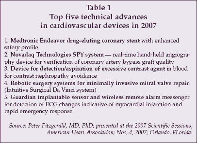 |
The first advance, safe formulations for DES devices, is an evolving trend that consists of a series of incremental changes in technology, beginning with devices such as the Endeavor and Endeavor Resolute stents from Medtronic (Minneapolis), that are now available outside the U.S. (the Endeavor in early November receiving an expert panel's positive recommendation for FDA approval).
Other next-generation stents that are expected to further enhance DES safety include the Xience V stent from Abbott Vascular (Abbott Park, Illinois), which will undergo a PMA review by the FDA in late November, and bioabsorbable stents under development by Abbott Vascular, Biotronik (Berlin, Germany) and others.
Additional advances in development for coronary stenting include a small-vessel Sirolimus-eluting stent from Cardiomind (Sunnyvale, California) and new stents for ostial lesions.
More from the top five
Another of the top five developments cited by Fitzgerald is new imaging technology for guidance of cardiovascular bypass surgery.
The SPY system from Novadaq Technologies (Mississauga, Ontario) is a cell-phone sized fluorescent imager that allows the cardiovascular surgeon to acquire an in situ angiogram during open-heart procedures, enabling the quality of bypass grafts to be verified in real time before the chest is closed.
A study performed with the system found that 8.2% of grafts that had been deemed satisfactory by visual inspection were defective by in-situ imaging. The SPY system is FDA-cleared, and U.S. Medicare reimbursement for use of the system in cardiac surgery became available in October 2007.
Other key advances cited by Fitzgerald include a new sensor and aspiration catheter for controlling the concentration of contrast agents during an angiogram to minimize contrast nephropathy, new surgical robotics and simulation technologies that provide improved consistency in cardiovascular surgery, and a new early detection system for myocardial infarction, the Guardian System, that can be implanted with a pacemaker to detect dangerous arrhythmias and automatically signal an emergency response team.
Advances in Imaging
A major area of focus at the AHA sessions was advances in non-invasive and invasive imaging modalities that are revolutionizing the diagnosis of cardiovascular disease and enabling more effective therapy.
CT imaging has dramatically improved with the advent of 64-slice systems, and is expected to become even more valuable as 256-slice systems enter the market.
At the AHA sessions, results of a study comparing 64-slice CT imaging to coronary angiography, the Core-64 trial, were presented by Julie Miller, MD, of Johns Hopkins University School of Medicine (Baltimore). The Core-64 trial involved nine centers in seven countries, and evaluated 291 patients who had both imaging procedures performed. A total of 8% of the patients initially enrolled in Core-64 were excluded, due to high levels of coronary calcium.
The study showed that multi-slice CT has a similar power to predict the need for revascularization as conventional angiography, with a sensitivity of 85% and specificity of 90% compared to angiography. However, CT could not accurately determine which individual vessel would require revascularization.
Miller said she believes the results could change the paradigm for diagnosis of coronary artery disease, with multi-slice CT providing a viable alternative to cardiac stress testing and nuclear imaging for initial work-up of at-risk patients, and eliminating unnecessary angiograms.
Leading suppliers of multi-slice CT systems include GE Healthcare (Milwaukee, Wisconsin), Philips Medical Systems (Best, The Netherlands), Siemens Medical Solutions (Erlangen, Germany) and Toshiba Medical Systems (Tokyo).
At the scientific sessions, Toshiba introduced an automated multi-slice CT analysis program, phaseXact, that selects the optimal phase of the CT signal for analysis, thus helping to streamline workflow and reduce interpretation time. These are important factors as the use of multi-slice CT in initial work-up of patients with suspected coronary artery disease expands.
However, a question often unasked at the AHA sessions is how often will these advancements reach a broad spectrum of patients, given their sophistication and high price? While the studies concerning these advances are impressive, their cost tends to emphasize the gap between those whose insurance will pay for them and those who have no insurance and/or no access to facilities providing these modalities.
Cardiovascular getting more MRI-ized
Another non-invasive imaging modality highlighted at the AHA sessions was MRI, including new targeted contrast agents that hold promise for improved characterization of arterial plaque.
Esad Vucic, MD, of Mt. Sinai School of Medicine (New York) described studies with p947, a new contrast agent from Guerbet (Villepinte, France) that targets matrix metalloproteinases which are known to occur in atherosclerotic plaque. Imaging with P947 provides a significant enhancement of high-risk plaques compared to control preparations that lack targeting.
P947 is not yet available in the U.S.
Another new development-stage MRI agent, BMS753951, was described at the sessions by Simon Robinson of BMS Medical Imaging (North Billerica, Massachusetts). The agent is a gadopentetate dimeglumine (Gd-DTPA) preparation that shows preferential uptake in arterial tissue. The uptake is associated with elastin fibers in arterial tissue and is highly specific, with the lungs being the only other site where significant accumulation is noted.
The agent provides at least 10-fold contrast of arterial tissue over other tissues except in lung, where the contrast ratio drops to about three-fold. The goal with targeted MRI agents is to noninvasively detect areas of vulnerable plaque, as indicated by a high concentration of markers such as MMP or macrophages. However, MRI still lacks adequate spatial resolution to equal the ability of CT to characterize arterial anatomy.
Furthermore, CT contrast agents now in development, such as N1177 from NanoScan Imaging (Landsdale, Pennsylvania), also may allow plaque characterization via molecular targeting of markers associated with vulnerable plaque such as macrophages.
Ultrasound is heard from
Ultrasound imaging is also becoming an increasingly important modality in cardiology, both for noninvasive imaging including the use of contrast agents as well as invasive imaging for both diagnosis and procedure guidance.
David Sahn of the Oregon Health and Science University (Portland, Oregon) described a new forward-looking ultrasound intracardiac imaging catheter under development with joint funding from GE Healthcare and St. Jude Medical (St. Paul, Minnesota). The device, manufactured by the Irvine Biomedical division of St. Jude, provides very high spatial resolution using near field ultrasound techniques, and includes a 24-element linear capacitive micromachined ultrasonic transducer (CMUT) array at the tip.
The catheter is being employed in guidance of ablation procedures for treatment of cardiac arrhythmias, using both high-intensity ultrasound (HIFU) and laser ablation devices. The catheter provides real-time feedback on the effectiveness of ablation, and is proving particularly useful as cardiac ablation techniques increasingly employ anatomical (rather than electrical) landmarks.
Acusphere (Watertown, Massachusetts) and POINT Biomedical (San Carlos, California) are two of the leading developers of ultrasound contrast agents for use in cardiology. Results from two studies using the Acusphere Imagify perflubutane polymer microsphere agent for myocardial perfusion imaging, the Phase 3 Real Time Assessment of Myocardial Infusion (RAMP-1) and RAMP-2 studies, were presented at the sessions.
As discussed by Paul Grayburn, MD, of Baylor University Medical Center (Houston, Texas), ultrasound agents, which typically consist of microbubbles that produce a high return signal when exposed to ultrasonic waves, can be used not only in diagnostic applications but also as delivery vehicles for targeted therapy. The results of the RAMP studies, which compared ultrasound perfusion imaging with Imagify to conventional nuclear (SPECT) imaging, showed that ultrasound gives equivalent specificity, and that sensitivity is somewhat higher.
POINT Biomedical's CARDIOsphere agent, which is comprised of echogenic bilayer microspheres (bioSpheres) filled with nitrogen, has also demonstrated sensitivity and specificity that are clinically equivalent to nuclear imaging, according to Grayburn.
Advantages of ultrasound perfusion compared to nuclear imaging include the wide availability of ultrasound equipment, shorter procedure time, lower cost, no exposure to radioactivity, and elimination of hazardous waste disposal issues.
Ultrasound with perfusion contrast
According to developers, roughly 10 million nuclear stress imaging procedures were performed in the U.S. in 2002, and the number of procedures is expected to grow at 7% annually over the next several years. The existing worldwide market for radioisotope nuclear imaging agents is estimated at $1 billion annually, creating a significant market opportunity for ultrasound perfusion contrast agents.
Potentially, the market could expand since ultrasound imaging can and is routinely performed in physician office and clinic settings, making it readily accessible to patients, whereas nuclear imaging is generally restricted to hospitals and large imaging centers.
In addition, there is the potential to extend the applications of ultrasound imaging to include molecular targeting of contrast agents to detect vulnerable plaque, monitor angiogenesis, and assess other important characteristics of vascular tissue.
For example, recent studies by Flordeliza Villanueva, MD, at the University of Pittsburgh Medical Center, have demonstrated ischemic memory imaging using ultrasound contrast, i.e., detection of a region of tissue that has experienced prior ischemia. The method could be used to assess the degree of re-perfusion following an infarction, or to examine a patient who had experienced an episode of chest pain which resolved to determine if an ischemic event occurred.
Ultrasound gets 'black box' treatment
One possible barrier to adoption of new ultrasound contrast agents is a recent black box label warning mandated by the FDA for existing agents such as GE Medical's Optison and Definity from Bristol-Myers Squibb.
The FDA required the label change in October 2007, due to reports of patient deaths and serious cardiovascular reactions in patients who received the agents. So far, ultrasound contrast agents are used in a small number of procedures, mainly in cases where non-contrast imaging does not provide a definitive result.
In a discussion of the black box warning at the AHA sessions, Grayburn strongly disagreed with the FDA decision, since it was based on reports of four deaths that were not definitively attributable to the agent used. Grayburn urged physicians to petition the FDA to dispute the decision and argue against any restriction on the use of ultrasound contrast.
In the RAMP studies, only one significant adverse reaction (a hypersensitivity reaction) was observed in more than 650 patients who received Imagify. On the other hand, SonoVue, an ultrasound contrast agent from Bracco International B.V. (Amsterdam, the Netherlands) used in Europe, was suspended from use in echocardiographic imaging procedures by the European Agency for Evaluation of Medicinal Products (EMEA; London) in 2004.
Another barrier to adoption of ultrasound imaging as a replacement for nuclear imaging in cardiac perfusion testing is the attractive reimbursement structure for nuclear imaging.
According to suppliers of nuclear imaging equipment, the modality ranks as the most profitable of all imaging technologies for physicians. Reimbursement from Medicare for a nuclear scan is approximately $800, and vendors can offer physicians leasing programs that allow realization of net income of about $120,000 per year when performing only six scans per week.
New ultrasound instrumentation
New developments in ultrasound instrumentation were exhibited at the AHA conference by Siemens Medical and GE Healthcare.
Siemens introduced the Acuson P50, a new entry in the hand-carried ultrasound segment developed via a partnership with Terason (Burlington, Massachusetts).
The system consists of an Apple MacBook laptop computer mounted on an ultrasound platform from Terason, weighs 12 pounds, provides two hour battery life, and has a base price of approximately $40,000. It is aimed at applications in emergency medicine, including use in ambulances and other emergency response settings, as well as private practice.
Although the P50 does not provide equivalent image quality to Siemens' top-of-the-line Sequoia ultrasound system, it offers more analytical features. Siemens also offers a palmtop ultrasound system, the P10, weighing 1.6 pounds with a one-hour battery life priced at under $10,000.
GE Healthcare introduced the Vivid S6 cardiovascular ultrasound system, a mobile system designed for use in a wide range of settings including the hospital, clinic, operating room, and physician's office. The Vivid 6 provides analytical features such as Tissue Velocity Imaging and Tissue Synchronization Imaging, along with stress echo and transesophageal echo capabilities.
Heading to the web
A new system for non-invasive early detection of coronary ischemia was exhibited at the AHA conference by Premier Heart (Port Washington, New York). The 3DMP system is a web-based two-lead resting ECG analyzer used by physicians for quantitative detection of ischemia in early-stage coronary artery disease, i.e., when coronary artery narrowing is only 40-50%.
The system relies on a proprietary signal analysis technique developed by Premier that detects stress and strain properties of the heart which are related to coronary artery abnormalities.
The technology was developed using data from a study involving 28,000 patients with known coronary artery disease and 7,000 normal individuals. Good correlation has been observed between the 3DMP results and test results obtained using other modalities such as intravascular ultrasound, coronary angiography, and CT angiography.
The system has received FDA 510(k) clearance and is priced at $29,000. Premier believes the 3DMP system will not only prove attractive to physicians because of its ability to non-invasively detect individuals at risk of a myocardial infarction well in advance of an event, but also because of the attractive reimbursement of $200 to $300 per exam, which is significantly higher than for a standard ECG exam because the 3DMP qualifies as a signal-averaged ECG analyzer.
Continued Expansion of PCI
A number of advances in device technology for percutaneous interventional treatment of heart disease were also reported at the AHA sessions.
The controversy over DES safety continues, although new data discussed at the conference indicates that previously published results — such as the SCAAR registry data, which indicated excess mortality for patients receiving DES vs. BMS) — do not pertain to current practice.
Specifically, as discussed by David Holmes, MD, of Mayo Clinic (Rochester, Minnesota) at a session on off-label use of DES devices, the initial conclusions from the Swedish SCAAR registry were based on data from patients treated in 2003 to 2004, whereas more recent data from SCAAR, reflecting long-term follow-up of patients treated in 2005, indicates that DES devicess are superior to BMS with respect to the endpont of mortality.
Holmes said it is now clear that the quality of stent implantation is critical for DES, including the need for high-pressure inflation, in order to achieve optimal long-term safety.
In addition, as indicated by other studies presented at the AHA sessions, avoiding use of DES in long lesions (>31.5 mm) may provide a significant improvement in safety, since late stent thrombosis rates in long lesions are up to seven-fold higher (3.5% vs. 0.5%) than in shorter lesions.
Holmes also referenced new data about to be published which will indicate that late stent thrombosis, the major safety issue for DES, also has been found to occur with BMS.
DES: safer than previously thought
Overall, the most recent results from clinical trials and registries indicate that DES may be safer than believed about a year ago when the results of SCAAR and other studies were first announced, possibly leading to a stabilization in utilization rates, if not an uptick in use.
While utilization has fallen dramatically in some settings (i.e., Sweden, where the DES utilization rate fell from 60% to 20%), rates of DES use remain quite high in many U.S. centers.
For example, Holmes quoted 80% DES use at the Mayo Clinic, while Cindy Grines, MD, quoted a rate of 70% at Royal Oak Medical Center (Detroit, Michigan).
But the first and second generation modalities of DES devices may be relatively short-lived as an increasing number of researchers and physicians are looking to bioabsorbable stents as an alternative to devices that remain in the body. A report on Biotronik's product in this area indicated relatively rapid absorption of the stent and no adverse events.
The amount of evidence needed for these devices and how soon they will reach the market are very large question marks, but they are likely to be common in the next decade.
Addressing heart defects
New developments in management of patients with heart defects such as Patent Foramen Ovale (PFO) and Atrial Septal Defects (ASDs) were another topic highlighted at the AHA sessions. The market for devices used in heart defect repair has exhibited substantial growth recently, as shown in Table 2.
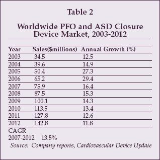 |
Key suppliers of PFO and ASD closure devices include AGA Medical (Plymouth, Minnesota), NMT Medical (Boston), Cardia (Eagan, Minnesota), St. Jude Medical and W.L. Gore (Flagstaff, Arizona). At present, only AGA Medical, NMT and W.L. Gore market PFO and ASD closure devices in the U.S.
The potential market, according to suppliers for cardiac septal repair implant devices, is more than 4.5 million procedures for migraine headache and about 750,000 procedures annually for stroke and TIA. The worldwide market for devices used to repair congenital heart defects, such as Atrial Septal Defects and Ventricular Septal Defects, is about 30,000 procedures.
AGA shipped 60,000 devices of all kinds in 2006 worldwide, and cumulative shipments totaled 250,000 as of August 2007, while about 23,000 PFOs have been closed worldwide using NMT's devices.
As discussed by Stephan Windecker, MD, of University Hospital Bern (Bern, Switzerland), use of closure devices for stroke prevention is well established, reducing the risk of cryptogenic stroke and transient ischemic attack from 5.2% for patients treated with medical therapy to 1.2% for patients treated with mechanical closure. In addition, the risk of bleeding associated with medical therapy is reduced with mechanical closure.
Applications in migraine headache prevention are less well established, since results of the first Migraine Intervention with STARFlex Technology (MIST) trial which evaluated the NMT Medical STARFlex closure device showed no difference in the number of patients experiencing total cessation of migraine in the treatment group vs. controls.
However, as discussed by Aaron Kaplan, MD, of Dartmouth-Hitchcock Medical Center (Lebanon, New Hampshire), if the effect of closure is assessed in terms of the number of patients achieving a 50% reduction in headache symptoms, as is the case with medical therapy, a more significant response rate is observed.
In October, NMT initiated the MIST II trial, employing the new BioSTAR bioabsorbable closure device, which offers improved sealing and also has the advantage of not leaving a permanent implant in the patient. More than 20 million patients are potential candidates for PFO closure in the U.S. alone, according to Kaplan.
Other suppliers developing devices for PFO and ASD closure include Cierra (Redwood City, Calilfornia) Sutura (Fountain Valley, California); Coherex Medical (Salt Lake City); NDC Inc. (Fremont, California), developing the SeptRx device; Coaptus Medical (Redmond, Washington), developing a radiofrequency closure device; and Swissimplant AG (Solothurn, Switzerland), developing the Solysafe Septal Occluder.
Devices for ablation
Another segment of the interventional device market highlighted at the AHA sessions is ablation devices for treatment of cardiac arrhythmia. Advances were discussed not only in ablation technology, but also in imaging modalities for procedure guidance.
Yasuo Okumura of Mayo Clinic (Rochester, Minnesota) discussed an evaluation of the new CartoSound Module from Biosense Webster, a unit of Johnson & Johnson (New Brunswick, New Jersey).
The CartoSound system, which interfaces to the Siemens Acuson Sequoia and Cypress intracoronary ultrasound imaging systems to provide guidance information for ablation procedures, was compared to the CartoMerge guidance system, which relies on creation of electroanatomical maps that are registered with CT imaging to heart anatomy.
Okumura found that CartoSound imaging is more accurate than CartoMerge imaging, providing a positional accuracy of 1 mm compared to 2.5 mm with CartoMerge. Such advances in guidance could prove important in efforts to improve the efficacy of coronary ablation of cardiac arrhythmias. With existing techniques, the success rate for arrhythmia ablation is only about 50%, according to suppliers of ablation devices, leading to a number of development efforts aimed at more effective approaches.
Emulating Cox-Maze
One new technology recently launched in the U.S. market, the VisiTrax system from NContact Surgical (Morrisville, North Carolina), employs a linear RF electrode array that is held in contact with the heart tissue using an integrated suction cup. Rather than creating individual ablation foci as is done with an RF ablation catheter, the VisiTrax device allows any ablation pattern to be created, including emulation of patterns used in the proven Cox-Maze surgical ablation procedure.
The procedure is performed on the beating heart, and can be implemented either via open surgery or in a minimally invasive manner using port-access techniques. The VisiTrax received FDA clearance in September, and is now being introduced in the U.S.
Another new ablation technology, described by Arjun Sharma, MD of Regional Cardiology (Sacramento, California), is under development by CyberHeart (Menlo Park, California). The company was founded in 2005 by Thomas Fogarty, MD, and Roderick Young.
The CyberHeart system employs non-invasive stereotactic radiation for cardiac ablation, using the CyberKnife radiotherapy system from Accuray (Sunnyvale, California) as the radiation source.
Stereotactic radiation has already been employed for a variety of ablation procedures, including inducing A-V block.
The initial application in cardiac ablation being addressed by CyberHeart is pulmonary vein ablation for treatment of atrial fibrillation. The system provides highly accurate control of ablation patterns, using sensing techniques to compensate for body motion and respiration-related movement.
Beam position can be adjusted to avoid ablation of coronary arteries and other vital structures, and both pulmonary veins can be ablated in a single procedure. The left atrial appendage can also be ablated if desired, which is difficult to accomplish using catheter ablation. Less than 1% of the radiation dose is delivered to the skin using stereotactic radiation, allowing most of the dose to be delivered to the target tissue.
Although at present trials are being performed using metal clips as fiducials to guide the CyberKnife, techniques are being developed that will eliminate that requirement, making the procedure completely noninvasive. At present, procedure time is about one hour. One drawback of the procedure is that the therapeutic effect does not occur immediately, presumably because of the gradual response of tissue that is typical in radiation treatment. Between eight and 10 weeks are required for complete response. However, the non-invasive nature of the CyberHeart system could allow use of cardiac ablation to expand significantly.
At present, according to suppliers, about 19,000 EP ablation procedures are performed annually in the U.S. out of an estimated two million to four million patients with nodal-related arrhythmias.
Transcatheter heart valves
Transcatheter heart valves represent an emerging market opportunity for device suppliers including existing manufacturers of artificial valves as well as development-stage companies.
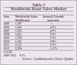
As shown in Table 3, the worldwide market for heart valves totaled over $1.4 billion in 2006, and is forecast to grow substantially due to the adoption of transcatheter devices, which are expected to begin to achieve significant market penetration beginning in 2009.
 |
At present, heart valve repair is performed on a small fraction of the patients who can potentially benefit. Suppliers estimate that only about 140,000 of the approximately 525,000 patients with severe valve disease, including those with mitral regurgitation, aortic stenosis, and pulmonary valve disorders, are treated at present, mainly because of the invasive nature of valve surgery. Transcatheter valve technology can potentially enable more patients to be treated, including those who are unable to undergo surgery as well as those with significant disease who are presently not at a sufficiently advanced stage to warrant costly open surgery.
As discussed by Helene Eltchaninoff, MD, of Rouen, France, at the AHA sessions, about 30% of patients with severe aortic stenosis are candidates for percutaneous aortic valve therapy, technically the least difficult of the heart valves to repair via percutaneous techniques.
Leaders in the race to develop transcatheter heart valves include Edwards Lifesciences (Irvine, California), CoreValve (Irvine, California), and EValve (Menlo Park, California), and other companies developing transcatheter heart valve repair devices include Ample Medical (Foster City, California), St. Jude Medical, Cardiac Dimensions (Kirkland, Washington), Viacor (Wilmington, Massachusetts), Mitralign (Tewksbury, Massachusetts), QuantumCor (San Clemente, California), and Myocor (Maple Grove, Minnesota).
To date, based on figures quoted by Eltchaninoff, 550 patients have received implants of the Edwards Sapien aortic valve, and 350 patients have received implants of the CoreValve aortic ReValving System in prospective clinical trials. Both devices employ biologically derived pericardium to form the valve leaflets, along with an expandable metal frame to form the valve body. The Edwards device received CE marking in September 2007. Short-term success rates have been high for both devices: procedural success for the CoreValve was quoted at 92% by Eltchaninoff, and follow-up with the Edwards device has now been tracked out to four years.
Mitral valve repair using percutaneous technologies is proving more challenging, however. As discussed by Peter Block, MD, Emory University School of Medicine (Atlanta), clinical studies with the Edwards Monarc device have resulted in a reduction in mitral regurgitation in 60% of treated patients. Other devices have shown similar rates of effectiveness.
For example, the Viacor PTMA device has achieved a 44% reduction in mitral regurgitation in clinical trials, while the CARILLON device from Cardiac Dimensions has achieved a 63% success rate for reduction of mitral regurgitation in the AMADEUS trial. Complete elimination of mitral regurgitation is probably not required, since, as discussed by Blasé Carabello, MD, of Baylor College of Medicine (Houston), patients can tolerate a one-third reversal of flow through the mitral valve without significant adverse consequences.
Overall, percutaneous therapy is proving successful at reducing mitral regurgitation by one grade in about 50% of patients.
Other devices in development for mitral regurgitation therapy, such as the Myocor Coapsys system, the PS3 device from Ample Medical, and the QuantumCor radiofrequency valve remodeling catheter, may achieve improved success rates. So far, percutaneous heart valve repair devices, particularly those designed for mitral valve repair, have not achieved success rates equivalent to those that can be obtained using open surgery.
However, as discussed by Donald Glower, MD, of Duke University Medical Center (Durham, North Carolina) at the AHA sessions, percutaneous valves may allow additional patient groups with valve disease to be treated, including those who are unable to undergo open surgery, and those with less severe disease who wish to avoid the risk and morbidity associated with surgery but can still realize an improvement in quality of life as a result of valve repair.
The market forecast for heart valves shown in Table 3 assumes that percutaneous therapy will enable an expansion of the addressable patient population for valve repair.