
BB&T Contributing Editor
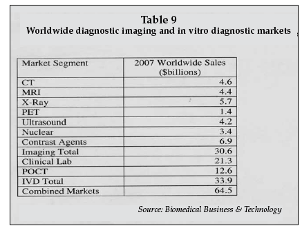
The integration of in vitro diagnostics and in vivo imaging is an emerging trend in diagnostics, exemplified by strategic initiatives from companies such as Siemens Healthcare (Munich, Germany). The combined worldwide markets for diagnostic imaging and in-vitro diagnostic products total-ed $64.5 billion in 2007, as shown in Table 8.
The in vitro diagnostics market is growing at a rate of 5%-6% a year while diagnostic imaging is growing at 2%-3% a year (both currency-adjusted), according to suppliers in those markets.
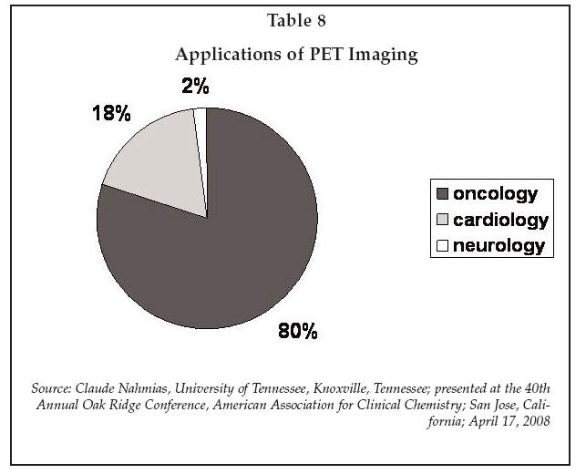
Molecular imaging is an example of an application segment where synergy exists between IVD and imaging, wherein a biomarker monitored in blood to detect disease or track progression can also be imaged to determine its spatial distribution in the body. At present, most molecular imaging is performed by positron emission tomography (PET), and is primarily employed in cancer diagnostics, as shown in Table 9.
As described by Claude Nahmias of the University of Tennessee (Knoxville) at the Oak Ridge Conference, molecular imaging allows in vivo detection of changes in tissue function, vs. morphological imaging which only detects changes in shape.
Detection of functional changes can enable earlier detection of disease, ac-cording to Nahmias, and molecular imaging can also identify targets for drug therapy. PET imaging, however, is limited by its low spatial resolution, making it difficult to precisely relate functional changes observed via molecular imaging to morphological changes detected by other modalities. Consequently, integrated PET/CT systems, and more recently PET/MR systems, have been introduced to provide high-resolution morphological information combined with functional information.
In cardiology, PET/CT systems are now used to compare metabolism to perfusion to assess myocardial viability and also to detect vulnerable plaque, necrosis and abnormal metabolism. In oncology, molecular imaging can be used to analyze tumor metabolism, DNA synthesis, and tissue differentiation.
However, as discussed by Nahmias, existing molecular imaging agents lack specificity. For example, fluorodeoxythymidine has been evaluated as a marker for cell proliferation via its correlation with DNA synthesis, but uptake in multiple organs limits its effectiveness.
More specific imaging probes are needed for molecular imaging to reach its full potential. However, development is likely to be slowed by the lengthy and costly regulatory approval process required for in vivo agents, and market acceptance will be impeded by the likely high cost of the agents.
Thomas Meade of Northwestern University (Evanston, Illinois) and recent founder of Ohmx Corporation (Evanston), a developer of handheld IVD biosensor devices, described new molecular imaging agents that demonstrate where the field may be headed. Meade has developed a family of molecular MR probes that take advantage of the high spatial resolution of MR and that are biochemically activated in vivo, giving distinct strong and weak relaxivity states.
Modulation between the two states is triggered by enzymatic processing of the agent, allowing spatially localized detection of specific enzyme activity, and by binding of intracellular messengers such as calcium and zinc, potentially enabling cell signaling mechanisms to be analyzed in vivo.
Meade has also developed iron oxide labels that provide enhanced sensitivity for MR contrast agents, enabling lower doses to be used compared to existing gadolinium-based agents.
In a second presentation dealing with molecular MR contrast agents, Alexander Guimaraes of Massachusetts General Hospital (Boston) described the use of magnetic nanoparticle agents that can be used to analyze tumor vascularity. The agents have potential applications in assessing the effect of anti-angiogenic agents such as VEGF inhibitors and rapamycin in renal cell and other cancers.
Experiments using the MR nanoparticle agent in mice show that microvascular density as measured by MR correlates with histological measurements, and that changes in vascularity as a result of treatment with antiangiogenic agents can be tracked.
Another presenter at the Oak Ridge Conference, James Wiskin of Techniscan Medical Systems (Salt Lake City, Utah), discussed advances in ultrasound imaging that may enable high-resolution molecular imaging to be performed with that modality, which is typically less costly and more widely available compared to MR.
Inverse scattering
The advances are based on unique inverse scattering mathematical algorithms developed by Techniscan for processing of ultrasound signals, and can enable resolution as high as 1 mm to be achieved. At that level, Wiskin said that information on tissue microstructure can be obtained, comparable to that provided by MR imaging.
The microstructural information can be used directly in some cases, for example in the detection of malignancy in breast images. Techniscan has already installed three systems in hospitals that have been used to image over 500 patients following conventional mammography, and shown that the Techniscan 3D ultrasound image can detect small tumors that are not visible via mammography.
The ultrasound method can potentially be enhanced through the use of various agents that selectively modify the acoustic properties of tissue at a cellular level such as microbubbles, liposomes, or perfluorocarbon nanoparticles along with ligands such as antibodies and peptides to provide molecular specificity.
A key advantage of the Techniscan technology is its ability to achieve millimeter-scale resolution at ultrasound frequencies of around 2 megahertz, where tissue penetration is very high, versus conventional ultrasound imaging which must use frequencies of 10 megahertz to achieve the same spatial resolution, at which penetration is very low.
For applications in breast cancer screening, the technology also offers the advantage of improved patient comfort during an exam compared to mammography, since the ultrasound image is obtained by suspending the breasts in a water bath, avoiding the sometimes painful mechanical compression required for x-ray mammography.
A new in vitro diagnostic technology for disease screening and risk assessment was described at the conference by Mickey Urdea, PhD, of Tethys Bioscience (Emeryville, California).
Tethys has developed a multi-marker assay, the PreDx diabetes test, to predict the risk of developing diabetes. The assay is to be introduced as a reference laboratory test in summer 2008. It measures a panel of protein biomarkers discovered in a study conducted in Finland and Sweden involving 9,000 subjects who were monitored over a five-year time frame for conversion to diabetes.
Diabetes risk high
A high percentage of the population is at risk of developing diabetes (estimated at 40 million to 60 million in the U.S.), based on existing risk assessment models, but at present the methods for predicting who will actually convert to diabetes are complicated and also do not allow tracking of the reduction of risk as a result of preventive therapy, such as metformin treatment.
An effective risk prediction test could have significant clinical value, since studies have shown that preventative treatment can reduce the incidence of conversion to diabetes in at-risk individuals by 25%-67%.
The Tethys test employs an ultrasensitive immunoassay system, Molecular Counting Technology, developed by Singulex (Alameda, California), providing a single number that can be used by a primary care physician to determine if preventive treatment is warranted.
Validation studies have shown that about 10% of the individuals who are candidates for the test, i.e. —those with a body mass index equal to or greater than 25 and are age 39 or older with a family history of diabetes — are at high risk of conversion. At that level, it would be practical to treat those who test positive.
The studies conducted by Tethys have shown a 500% increase in risk of conversion to diabetes for individuals who are positive by the PreDx test. In addition, a significant number of patients who are positive have normal levels of fasting plasma glucose, and would not be detected as diabetic by conventional screening methods. The Tethys assay will probably be priced at about the average cost for comparable genetic risk assessment tests such as those used to assess breast cancer risk, e.g., about $1,500.
A new test for assessment of the risk of prostate cancer recurrence after radical prostatectomy, the NADIA total PSA assay, was described in a poster presented at the conference by Jonathan McDermed of IRIS Diagnostics (Chatsworth, California).
About 70,000 radical prostatectomies are performed each year in the U.S., resulting in a cure in 80% of cases. Twenty percent of treated patients will have a recurrence, however, and require further treatment. Early, reliable detection of recurrence is desirable to enable additional treatment to be initiated when it is most effective, but existing PSA tests do not give reliable results at levels less than 0.01 ng/mL.
The NADIA assay, in contrast, has a sensitivity of 0.0005 ng/mL, enabling earlier detection of rising PSA levels after radical prostatectomy or cryotherapy. A study conducted by IRIS showed that, on average, the NADIA test detected a rising PSA 34 months before the total PSA value reached 0.1 ng/mL.
Advances in Analytical Technologies
A number of new analytical platform technologies were described at the conference that could provide a basis for next-generation diagnostic testing systems in the clinical laboratory.
One, under development by BioNanomatrix (Philadelphia), is based on the use of nanofluidic-enabled single-molecule imaging, and has potential applications in nucleic acid diagnostics.
BioNanomatrix has developed a fabrication technology that can produce fluidic channels with a diameter of only 10 nanometers that are several centimeters long. At that diameter, only a single nucleic acid molecule can pass through the channel at a time. Using high-resolution imaging, single molecules can be analyzed as they transit the channel. Electro-osmotic forces are used to drive the molecules through an array of parallel nanochannels, allowing thousands of molecules to be analyzed simultaneously. Up to 56,000 nanochannels can be fabricated on a chip the size of a typical microarray (1 square cm).
Applications include DNA sequencing, analysis of DNA-protein interactions, rapid measurement of FISH assays, and analysis of DNA methylation in cancer diagnostics. Materials cost for sequencing of an entire genome using the BioNanomatrix technology is estimated at $100. BioNanomatrix is collaborating with Complete Genomics (Sunnyvale, California) to develop applications in high-throughput gene sequencing.
Cell diagnostics is another growing segment of the clinical diagnostics market, with applications in detection of cancer metastasis, chemotherapy drug response, and cytology.
David Beebe of the University of Wisconsin (Madison) reported on developments in microfluidics technology with applications in clinical diagnostics. While microfluidics has been applied to biochemical screening and DNA sequencing, there has so far been no significant impact of the technology is cellular analysis. In part, the lack of applications is due to the complexity of microfluidics systems required for cell analysis.
Beebe described a new approach that uses tubeless microfluidics, which relies on surface tension in microdroplets to drive the movement of cell suspensions through an analytical microfluidic array. Depending on where a droplet containing a cell suspension is placed, and the size of the droplet, the direction of flow patterns can be controlled with high precision. In addition, cell/cell interactions via messenger molecules occur rapidly and in a controlled manner, and cells can be readily imaged using a variety of techniques including phase contrast.
Applications being developed by Beebe include tumor cell invasion assays, angiogenesis assays for drug discovery, and monitoring of tubule formation in cells exposed to drugs.The technology also may have applications in monitoring of drug susceptibility of cell biopsy samples, and in analysis of needle biopsy specimens.