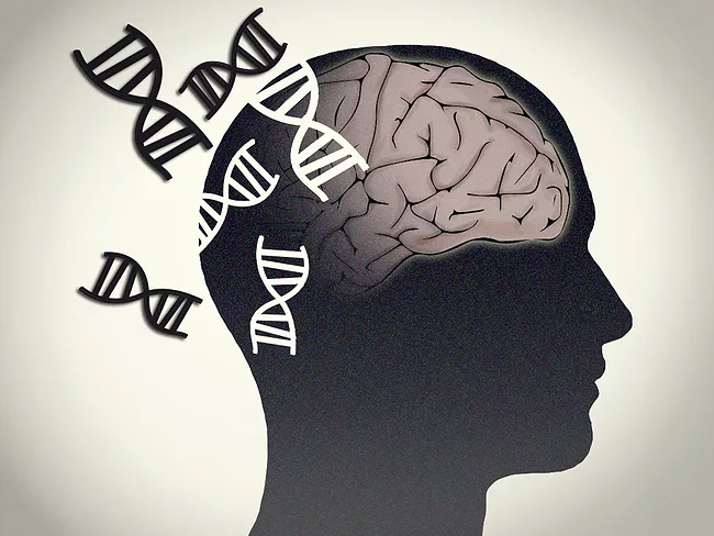
Genes that are switched on or off in the human brain differ between men and women. Moreover, these differences are not uniform. They vary across cortical regions and cell types. Scientists at the National Institute of Mental Health (NIMH) and the National Institute on Aging (NIA) used single-cell sequencing and unveiled distinct gene expression patterns regulated by hormones and sex chromosomes. This detailed map of the brain’s molecular biology shows how women and men switch on and off more than 3,000 brain genes differently and expands the catalogue of X chromosome genes that escape inactivation.
PerturbAI has emerged from stealth mode with the release of the world’s largest in vivo CRISPR atlas as described in a preprint on Biorxiv. The study profiled over 7.7 million cells from the brains of 74 mice with different cellular knockouts of 1,947 disease-associated genes. The San Francisco-based company’s Perturb-seq platform combines CRISPR perturbations with single nucleus RNA sequencing to look at gene expression.
A collaborative effort is trying to construct the cell atlas of the developing brains of humans and animal models. Using advanced single-cell and spatial technologies, they mapped how brain cell types emerge, diversify and organize over time, offering new insights into the origins of neurodevelopmental and psychiatric conditions. These breakthroughs in genomics and imaging will allow scientists to study complex developmental processes with high resolution.
A collaboration of scientists from the NIH Brain Initiative consortium has published eight simultaneous studies in Neuron, Cell, Cell Genomics, Cell Reports and Cell Reports Methods, with the results of the Armamentarium project, a new set of gene therapy tools for the research and treatment of human brain disorders. The methodology, based on genetic techniques, RNA detection, genomic enhancers and viral vectors, is designed to access different CNS cell types, neuronal and non-neuronal cells, with common and reproducible protocols now available for any laboratory.
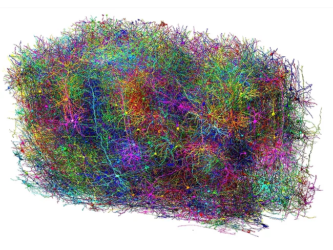
Scientists from the Machine Intelligence from Cortical Networks (MICRONS) consortium have published the microconnectome of a cubic millimeter of the mouse brain. This is the most complete map of this organ to date at nanometer resolution for a mammal. It not only contains the structure and connections of each and every cell in that volume of tissue, but is also linked to the neuronal activity of that portion of the CNS, linking anatomy and function in the same cells.
2024 saw the completion of several cellular-resolution brain maps, including the entire fly brain and a comprehensive connections map of a cubic centimeter of human brain. 2025 began with the addition of another important map. In the Jan. 1, 2025, issue of Nature, researchers from the Allen Institute presented a map of areas and cell types where aging most affected the mouse brain.
2024 saw the completion of several cellular-resolution brain maps, including the entire fly brain and a comprehensive connections map of a cubic centimeter of human brain. 2025 began with the addition of another important map. In the Jan. 1, 2025, issue of Nature, researchers from the Allen Institute presented a map of areas and cell types where aging most affected the mouse brain.
In the 1970s, scientists from several countries proposed to reconstruct, one by one, all the neurons in the brain as they appear under an electron microscope. They started with a small worm. Caenorhabditis elegans has only 302 neurons. It took 16 years. How much time would be required to repeat this arduous task for the 100 billion neurons in the human brain?
In the 1970s, scientists from several countries proposed to reconstruct, one by one, all the neurons in the brain as they appear under an electron microscope. They started with a small worm. Caenorhabditis elegans has only 302 neurons. It took 16 years. How much time would be required to repeat this arduous task for the 100 billion neurons in the human brain?
In the 1970s, scientists from several countries proposed to reconstruct, one by one, all the neurons in the brain as they appear under an electron microscope. They started with a small worm. Caenorhabditis elegans has only 302 neurons. It took 16 years. How much time would be required to repeat this arduous task for the 100 billion neurons in the human brain?